


Author:- Mr. Ritesh Sharma
As you must already know, an ECG machine showcases its results in the waveform patterns. These waveform patterns are P-wave, QRS complex, and T-wave. Within the QRS complex lies the R-wave. While the R Wave ECG is not as widely discussed as the other ECG waveforms, it is extremely crucial and has numerous clinical implications. It is essential to understand the R wave ECG to get a good understanding of the ECG waveforms in general. It gives you insights into different heart abnormalities as well.
In this blog, we will try to decode all the intricacies of the R Wave ECG. So, all healthcare professionals and general people alike will get worthy information from this blog.
What is an ECG?
An ECG is a non-invasive diagnostic tool that records the electrical activity of the heart over time using electrodes placed on the skin. These electrodes detect the electrical impulses generated by cardiac muscle depolarization and repolarization during each heartbeat. The resulting waveform, displayed graphically on paper or a monitor, comprises several distinct phases, each representing specific events in the cardiac cycle.
Anatomy of an ECG Waveform
The ECG waveform typically consists of several waves and intervals:
- P wave: Represents atrial depolarization, indicating the initiation of atrial contraction.
- QRS complex: Comprises the Q, R, and S waves, reflecting ventricular depolarization and contraction.
- T wave: Signifies ventricular repolarization, preparing the heart for the next cycle.
R Wave ECG
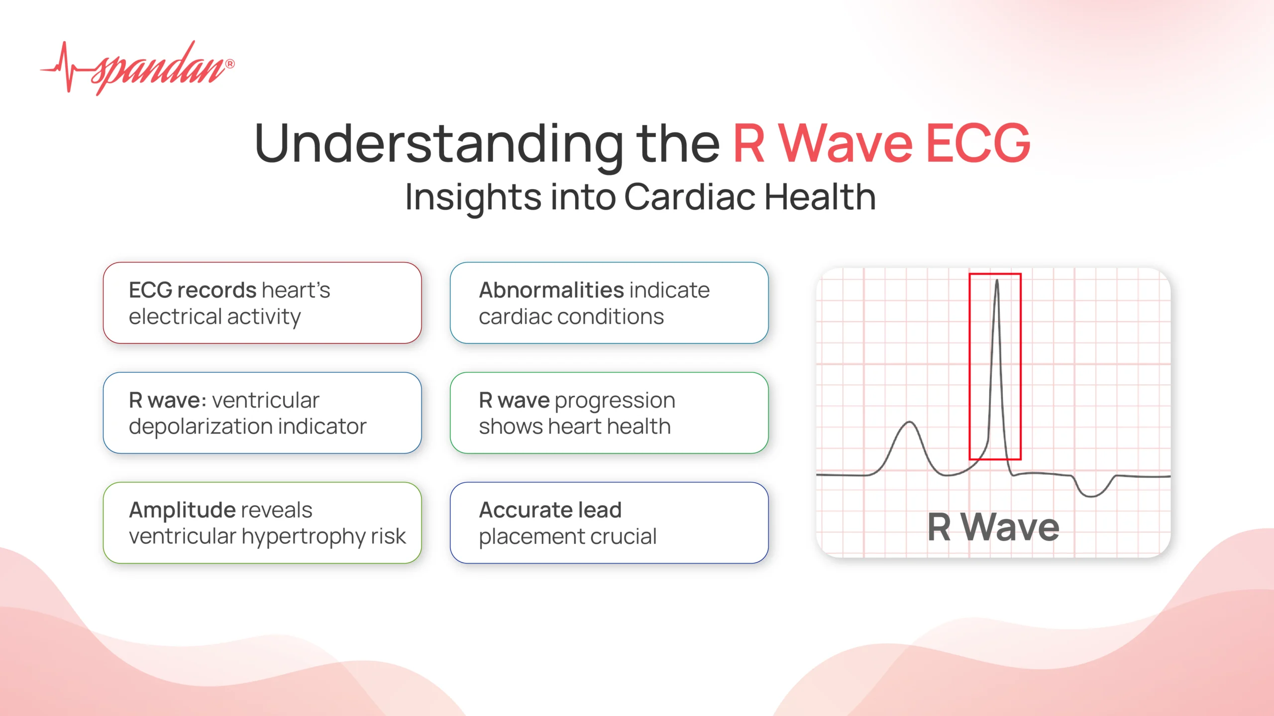
The R wave, a prominent peak within the QRS complex, marks the initial phase of ventricular depolarization. It signifies the rapid and coordinated contraction of the ventricles, essential for effective pumping of blood into the circulatory system. Clinically, the characteristics of the R wave provide valuable insights into various aspects of cardiac function:
- Amplitude: The height of the R wave correlates with the amount of electrical activity during ventricular depolarization. Higher amplitudes can indicate hypertrophy (enlargement) of the heart muscle, often seen in conditions like hypertension or chronic heart disease.
- Duration and Shape: The duration and morphology of the R wave can reveal abnormalities in conduction pathways or structural changes within the heart. For instance, a widened or distorted R wave may suggest conduction delays or myocardial infarction (heart attack).
- Lead Specificity: The appearance of the R wave varies across different ECG leads, offering insights into the heart’s orientation within the chest cavity and the specific areas of myocardium (heart muscle) involved in depolarization.
Clinical Applications
ECG analysis involving the R wave plays a crucial role in:
- Diagnosis: Identifying various cardiac conditions such as cardiac arrhythmias, ischemic heart disease, and electrolyte imbalances.
- Monitoring: Assessing the effectiveness of treatments and interventions aimed at managing cardiac disorders.
- Risk Stratification: Predicting the likelihood of future cardiac events based on ECG findings, such as in patients with coronary artery disease.
Interpretation Challenges and Considerations
While the R wave provides valuable diagnostic information, its interpretation requires consideration of several factors:
- Patient Variability: Normal variations in cardiac anatomy and physiology can influence the appearance of abnormal ECG waveforms, necessitating a nuanced approach to interpretation.
- Artifact and Noise: External factors such as patient movement or improper electrode placement can distort ECG tracings, affecting the accuracy of R wave analysis.
- Integration with Clinical Context: ECG findings must be interpreted in conjunction with patient history, symptoms, and other diagnostic tests to formulate accurate clinical decisions.
Advanced Techniques and Future Directions
Advancements in technology, such as computerized ECG analysis and artificial intelligence, are enhancing the accuracy and efficiency of R wave interpretation. These developments aim to:
- Automate Diagnosis: Streamline the identification of subtle abnormalities and improve diagnostic accuracy.
- Facilitate Remote Monitoring: Enable real-time ECG analysis and remote transmission of data for timely clinical intervention.
Understanding R Wave Progression
R wave progression refers to the gradual increase in the amplitude of the R wave from the right to the left chest leads in a standard 12-lead ECG. This pattern is expected as the electrical impulse travels through the heart, starting from the right ventricle and moving towards the left ventricle. Typically, in the precordial leads (V1 to V6), the R wave starts small in V1, grows progressively larger until reaching its peak in V4 or V5, and then slightly decreases in V6.
Clinical Significance of R Wave Progression
The pattern of R wave progression provides crucial information about the heart’s electrical and structural integrity:
- Normal R Wave Progression: In a healthy heart, the R wave progression should follow a predictable pattern. Any deviation from this pattern can indicate underlying cardiac abnormalities.
- Poor R Wave Progression: This term describes an abnormally slow increase or decrease in R wave amplitude across the precordial leads. Poor R wave progression can result from several conditions, including:
- Anterior Myocardial Infarction: Damage to the anterior wall of the heart can disrupt normal depolarization patterns, leading to diminished R wave progression.
- Left Ventricular Hypertrophy: Enlargement of the left ventricle can alter electrical pathways, affecting the R wave pattern.
- Chronic Lung Disease: Conditions like chronic obstructive pulmonary disease (COPD) can change the position of the heart within the chest, impacting R wave progression.
- Conduction Abnormalities: Issues such as left bundle branch block can interfere with the normal propagation of electrical impulses, disrupting the expected progression of the R wave.
- Clinical Implications: Recognizing abnormal R wave progression is critical for early detection and management of these conditions. It can prompt further diagnostic testing, such as echocardiography or cardiac MRI, to confirm the underlying pathology.
- Lead Placement and Technical Factors: Accurate lead placement is essential for reliable R wave progression assessment. Misplaced leads can mimic poor R wave progression, leading to potential misdiagnosis. Ensuring correct electrode placement and checking for technical issues is a fundamental step in ECG interpretation.
Case Studies and Examples
Examining specific cases can illustrate the importance of R wave progression in clinical practice:
- Case 1: Anterior Myocardial Infarction: A 55-year-old male presents with chest pain. His ECG shows poor R wave progression in leads V1 to V4, suggestive of an anterior myocardial infarction. Further evaluation with cardiac enzymes and angiography confirms significant blockage in the left anterior descending artery, necessitating prompt intervention.
- Case 2: COPD Patient: A 70-year-old female with a history of COPD undergoes routine ECG. Her R wave progression appears abnormal due to lung hyperinflation shifting the heart’s position. Awareness of her chronic condition helps avoid unnecessary cardiac investigations, focusing instead on managing her pulmonary disease.
In conclusion, the R wave ECG serves as a pivotal marker in assessing cardiac function and diagnosing various cardiovascular conditions. Its amplitude, duration, and morphology provide valuable insights into the electrical activity of the heart, guiding clinical decision-making and patient management. As technology continues to evolve, the role of the R wave ECG interpretation will undoubtedly remain central in the field of cardiovascular medicine, ensuring timely and accurate diagnosis for improved patient outcomes.