


Author:- Mr. Ritesh Sharma
Hypokalemia ECG is a condition which is characterized by low serum potassium levels. This condition is considered potentially life-threatening. More so detecting hypokalemia ECG is quite a daunting task. However, this prompt detection is crucial for the immediate start of the treatment for this supposedly hazardous condition.
Understanding the key signs of hypokalemia on an ECG can help healthcare providers make quick and accurate diagnoses, thereby preventing serious complications. This blog delves into the primary ECG features indicative of hypokalemia and the underlying mechanisms.
Understanding Hypokalemia ECG
Potassium is a vital electrolyte in the body, essential for various physiological functions, including maintaining the electrical excitability of cells, particularly in the heart. Normal serum potassium levels range from 3.5 to 5.0 mEq/L. Hypokalemia is defined as a serum potassium level below 3.5 mEq/L. It can result from various conditions such as excessive loss of potassium through urine or gastrointestinal tract, inadequate potassium intake, or shifts of potassium into cells.
Hypokalemia affects cardiac electrophysiology by altering the resting membrane potential and the repolarization phase of the cardiac cycle. These changes manifest as distinct patterns on an abnormal ECG, which, if recognized promptly, can lead to early intervention and treatment.
Key ECG Signs of Hypokalemia
- Flattened or Inverted T Waves
One of the earliest signs of hypokalemia on an ECG is the flattening or inversion of T waves. T waves represent ventricular repolarization. Potassium plays a critical role in this phase, and low levels disrupt the normal repolarization process, leading to flattened or inverted T waves. These changes are more prominent in the precordial leads, particularly V2 to V4. - Prominent U Waves
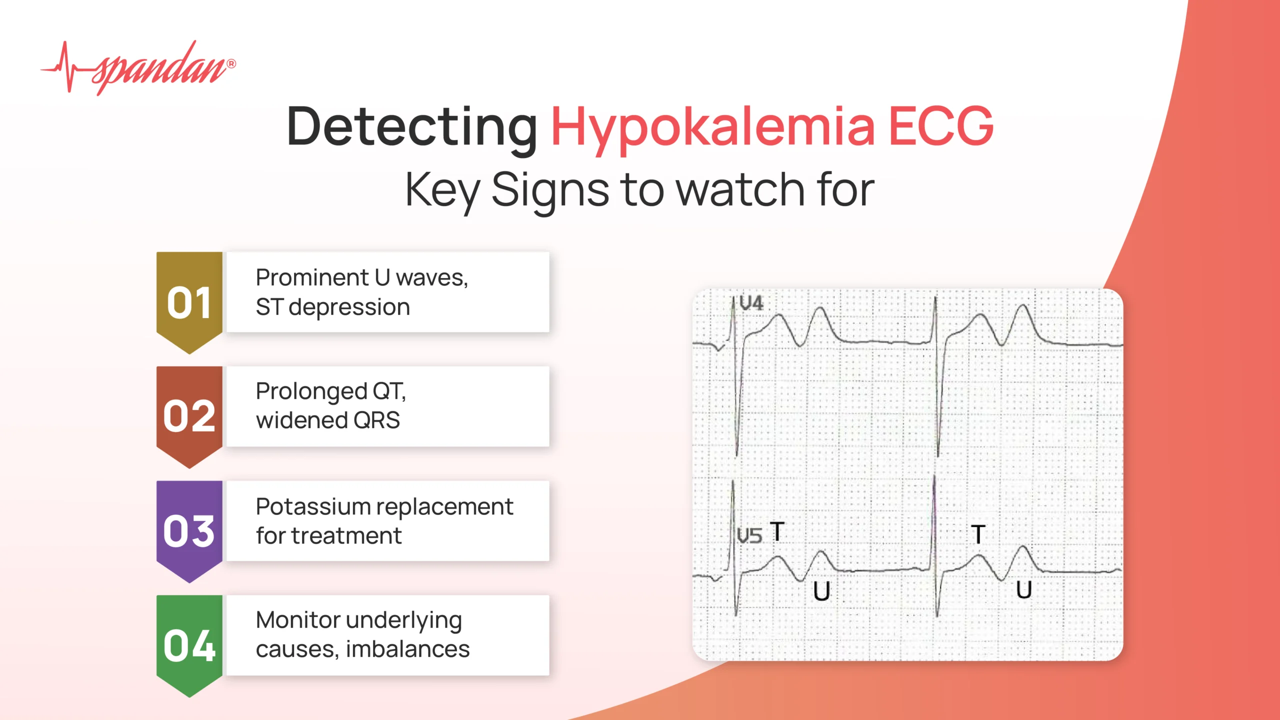
A hallmark of hypokalemia is the presence of prominent U waves. U waves are typically small, positive deflections following the T wave. In hypokalemia, these waves become more pronounced and can even surpass the amplitude of T waves in severe cases. Prominent U waves are best seen in the mid-precordial leads (V2 to V4). - ST Segment Depression
Hypokalemia can cause a depression of the ST segment, which represents the period when the ventricles are depolarized. ST segment depression in hypokalemia is often subtle but can be significant, particularly in the context of other ECG changes. It is important to differentiate this from ischemic causes of ST segment depression. - Prolonged QT Interval
Although the QT interval can appear prolonged due to the presence of a prominent U wave, it is crucial to differentiate the true QT interval from the QU interval (QT interval including the U wave). In hypokalemia, the true QT interval may be slightly prolonged due to delayed repolarization, but the apparent prolongation is often exaggerated by the prominent U wave. - Widening of the QRS Complex
In severe hypokalemia, the QRS complex can widen showcasing QRS complex abnormalities. This is due to the delayed depolarization of the ventricles. A QRS duration greater than 120 milliseconds should raise concern for severe hypokalemia or other electrolyte disturbances.
Mechanisms Behind ECG Changes in Hypokalemia
The ECG changes in hypokalemia are primarily due to the effect of low potassium levels on cardiac cell membrane potentials. Potassium ions play a crucial role in maintaining the resting membrane potential and facilitating repolarization. When potassium levels drop:
- The resting membrane potential becomes more negative (hyperpolarized), making it more difficult for cells to reach the threshold potential and depolarize.
- The repolarization process is delayed, leading to prolonged phase 3 of the action potential, which manifests as changes in the T wave and the appearance of U waves.
- The delayed repolarization can cause early afterdepolarizations, contributing to the risk of cardiac arrhythmias.
Clinical Significance and Management
- Potassium Replacement
The primary treatment is to correct the potassium deficit. This can be done orally for mild cases or intravenously for severe cases. The rate of intravenous potassium replacement must be carefully controlled to avoid hyperkalemia, which can also cause serious cardiac disturbances. - Addressing Underlying Causes
It is essential to identify and treat the underlying causes of hypokalemia. This may involve discontinuing or adjusting medications that cause potassium loss (e.g., diuretics), treating gastrointestinal losses, or addressing conditions that cause intracellular shifts of potassium. - Monitoring and Supportive Care
Continuous ECG monitoring is recommended for patients with significant hypokalemia, particularly if they are symptomatic or have severe electrolyte disturbances. Monitoring allows for early detection of arrhythmias and other cardiac complications. - Correction of Coexisting Electrolyte Imbalances
Hypokalemia often coexists with other electrolyte imbalances, such as hypomagnesemia, which can exacerbate cardiac instability. Correcting these imbalances is crucial for effective management.
In conclusion, Hypokalemia is a common electrolyte disturbance that can have serious cardiac implications. Recognizing the key ECG signs—such as flattened or inverted T waves, prominent U waves, ST segment depression, prolonged QT interval, and widened QRS complex—is essential for timely diagnosis and treatment.
Understanding the underlying mechanisms helps in appreciating the significance of these changes and guides appropriate clinical management. Prompt correction of potassium levels and addressing underlying causes can prevent life-threatening arrhythmias of different arrhythmia classifications and improve patient outcomes. Healthcare professionals must maintain a high index of suspicion for hypokalemia in patients with risk factors and be proficient in interpreting ECG changes to provide optimal care.