


Author:- Mr. Ritesh Sharma
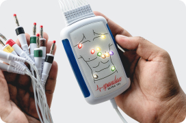
An ECG machine is an extremely important tool when it comes to cardiac care. This machine and its components are able to detect all kinds of different heart abnormalities ranging from heart palpitations to cardiac arrhythmias to heart attacks. One that’s an integral part of this machine besides ECG electrodes is ECG leads. Furthermore, the location of ECG leads is important as they capture the electrical impulses generated by the heart effectively. Correct placement of ECG leads is essential for accurate diagnosis and effective patient care.
Unfortunately, incorrect lead placement is a common issue that can lead to misdiagnosis and inappropriate treatment. This blog will discuss the common mistakes in ECG lead placement and provide tips on how to avoid them.
The Importance of Location of ECG leads
An ECG records the heart’s electrical activity from different angles, providing a comprehensive view of its function. Standard 12-lead ECGs use ten electrodes to capture this data, offering insights into heart rate, rhythm, and other critical factors. Incorrect placement of these leads can distort the recorded signals, leading to potential misinterpretations such as false positives or negatives for conditions like myocardial infarction, arrhythmias, and other cardiac abnormalities.
Common Mistakes in ECG Lead Placement
Incorrect Placement of Limb Leads: The four limb leads (RA, LA, RL, and LL) should be placed on the limbs as follows:
- RA (Right Arm): Right wrist or upper arm
- LA (Left Arm): Left wrist or upper arm
- RL (Right Leg): Right ankle or lower leg
- LL (Left Leg): Left ankle or lower leg
A common mistake is placing these leads on the torso, which can cause a deviation in the ECG readings known as limb lead reversal.
Misplacement of Precordial (Chest) Leads: The six precordial leads (V1-V6) must be positioned precisely on the chest to capture accurate heart activity:
- V1: Fourth intercostal space, right sternal border
- V2: Fourth intercostal space, left sternal border
- V3: Midway between V2 and V4
- V4: Fifth intercostal space, midclavicular line
- V5: Level with V4, left anterior axillary line
- V6: Level with V5, left midaxillary line
Common errors include incorrect intercostal space identification, misalignment with anatomical landmarks, and inconsistent spacing.
Reversal of Leads: Another frequent error is the reversal of leads, particularly the limb leads. This can produce an entirely abnormal ECG tracing, often leading to misdiagnosis. For instance, reversing the RA and LA leads can invert the P, QRS, and T waves, mimicking conditions like dextrocardia or anterior myocardial infarction.
Inconsistent Lead Placement: Variability in lead placement between different ECG recordings can make it challenging to compare results over time. This inconsistency can arise from different healthcare providers applying the leads in slightly different positions.
How to Avoid Common Mistakes for the Location of ECG Leads?
- Proper Training and Education: Regular training sessions and educational programs for healthcare providers on the correct placement of ECG leads can significantly reduce errors. This training should emphasize the anatomical landmarks and steps involved in precise lead placement.
- Use of Anatomical Landmarks: Ensure the correct identification of anatomical landmarks such as the sternal borders, intercostal spaces, midclavicular line, and axillary lines. Palpating these landmarks can help in accurate lead positioning.
- Standardization of Procedure: Developing a standardized protocol for ECG lead placement in healthcare settings can minimize variability. This protocol should be consistently followed and regularly reviewed to ensure compliance.
- Patient Positioning: The patient should be in a supine position with arms relaxed by their sides and legs uncrossed. This positioning helps in maintaining consistency and accuracy in lead placement.
- Double-Checking Leads: Before starting the ECG recording and figuring out the location of ECG leads, double-check the placement of all leads. This step includes verifying that each lead is correctly placed according to the standardized anatomical landmarks and ensuring no leads are reversed.
- Use of Diagrams and Checklists: Visual aids such as diagrams and checklists can be helpful reminders for proper lead placement. These tools can be placed in ECG rooms as quick references for healthcare providers.
- Regular Audits and Feedback: Conducting regular audits of ECG lead placements and providing feedback can help identify common mistakes and areas for improvement. This continuous feedback loop can enhance the accuracy of lead placement over time.
Advanced Techniques and Tools
- Color-Coded Leads: Using color-coded leads can help in quickly identifying and correctly placing each lead. This system reduces the likelihood of lead reversal and misplacement.
- Automated ECG Machines: Some advanced ECG machines have built-in prompts and error-checking mechanisms that alert the user if leads are improperly placed or if there is an issue with the connection.
- Simulation Training: Utilizing simulation training can provide hands-on experience without the risk of harm to patients. These simulations can mimic various scenarios and help healthcare providers practice and perfect their lead placement skills.
- Patient-Specific Adjustments: In cases where patients have unique anatomical features or are unable to lie flat, adjustments should be made with careful consideration. Documenting these adjustments ensures consistency in future recordings.
The accurate location of ECG leads is crucial for the correct interpretation of cardiac activity. Common mistakes, such as incorrect placement of limb and precordial leads, lead reversal, and inconsistent placement, can significantly impact the accuracy of an ECG. By adhering to proper training, using anatomical landmarks, standardizing procedures, and employing advanced tools and techniques, healthcare providers can minimize errors and improve the quality of ECG recordings.
Regular audits, feedback, and continuous education are essential to maintaining high standards in ECG lead placement. With these practices in place, the reliability of ECG as a diagnostic tool can be significantly enhanced, leading to better patient outcomes and more effective cardiac care.