


Author:- Mr. Ritesh Sharma
The human heart has an intricate electrical system. This electrical system helps orchestrate the conduction system of the heart. Through this conduction system, the human heart contracts and relaxes and ensures it pumps blood to all parts of the body. Without the seamless performance of the conduction system of the human heart, the circulatory system won’t work properly and all parts of the body will not receive enough oxygen and nutrients to function.
Therefore, the conduction system of the heart is of utmost importance when speaking about different facets of the heart. The contraction of the heart muscle is initiated by the natural pacemaker of the heart and it gets complete and it is completed by Purkinje fibers where the contraction is coordinated.
In this blog, we will dive into various aspects of the conduction system of the heart and how it works in symphony with the chambers of the heart to coordinate the contraction of the heart muscle ultimately leading to circulation of blood to different parts of the body. So, whether you belong to the general audience or the category of clinicians, you will find worthy education and enlightenment from this blog.
Anatomy of the Conduction System of the Heart
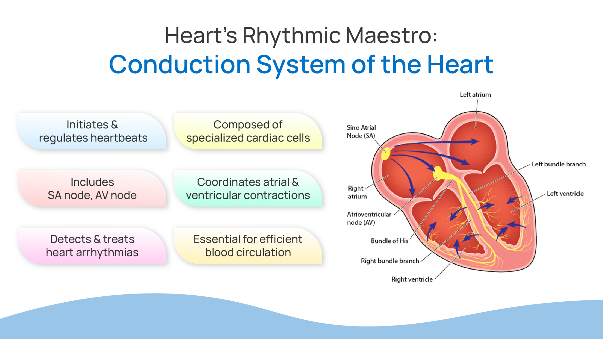
The conduction system of the heart is composed of specialized cardiac muscle cells capable of generating and conducting electrical impulses. The main components include the sinoatrial (SA) node, atrioventricular (AV) node, bundle of His, bundle branches, and Purkinje fibers. Each of these elements plays a crucial role in maintaining the heart’s rhythm.
- Sinoatrial (SA) Node: Often referred to as the heart’s natural pacemaker, the SA node is located in the right atrium near the superior vena cava. It initiates the electrical impulses that set the pace for the heart rate. The SA node generates impulses at a rate of about 60-100 beats per minute under normal conditions, ensuring that the heart beats in a regular rhythm.
- Atrioventricular (AV) Node: Situated at the junction of the atria and ventricles, the AV node serves as a critical gateway for electrical impulses. It delays the transmission of these impulses to the ventricles, allowing the atria to complete their contraction and the ventricles to fill with blood. This delay is essential for maintaining the coordinated contraction and efficient pumping action of the heart.
- Bundle of His: This collection of heart muscle cells is responsible for transmitting electrical impulses from the AV node to the ventricles. The Bundle of His bifurcates into the right and left bundle branches, which run along the interventricular septum.
- Bundle Branches: These branches further divide into smaller fibers that extend throughout the ventricles. The right bundle branch conducts impulses to the right ventricle, while the left bundle branch does so to the left ventricle. This branching network ensures that the electrical signal is distributed evenly and rapidly across the ventricular muscle.
- Purkinje Fibers: These fibers are the terminal ends of the conduction system. They spread throughout the ventricles and ensure that the electrical impulse reaches every part of the ventricular muscle, resulting in a coordinated contraction.
How does the electrical journey of the heart work?
The primary function of the conduction system of the heart is to ensure that the heart beats in a coordinated and efficient manner. Here’s a step-by-step breakdown of how the electrical impulses travel through the heart:
- Initiation at the SA Node: The process begins at the SA node, where an electrical impulse is generated. This impulse spreads rapidly through the walls of the right atrium and then to the left atrium, causing both atria to contract simultaneously and push blood into the ventricles.
- Transmission to the AV Node: The impulse then reaches the AV node, which delays it slightly. This delay ensures that the ventricles have enough time to fill with blood from the atria before they contract.
- Passage through the Bundle of His and Bundle Branches: From the AV node, the impulse travels down the Bundle of His, which splits into the right and left bundle branches. These branches conduct the impulse to the respective ventricles.
- Distribution via Purkinje Fibers: Finally, the impulse reaches the Purkinje fibers, which distribute it throughout the ventricles. This triggers a synchronized contraction of the ventricular muscles, propelling blood out of the heart – the right ventricle sends blood to the lungs, and the left ventricle sends blood to the rest of the body.
Heart Rhythms and Disorders
The disruption in the conduction system of the heart can cause several disorders. These disorders are cardiac arrhythmias of different arrhythmia classifications. In this, the rhythm of the heart is irregular, i.e. it either beats too fast, too slow, or irregularly. Let’s learn more about these disorders in detail:
- Bradycardia: This condition is characterized by a slow heart rate, typically below 60 beats per minute. It can result from issues with the SA node or disruptions in the conduction pathways. Severe bradycardia can lead to fatigue, dizziness, and fainting.
- Tachycardia: Opposite to bradycardia, tachycardia involves a fast heart rate, usually over 100 beats per minute. This can be caused by abnormal electrical signals originating in the atria (supraventricular tachycardia) or ventricles (ventricular tachycardia). Symptoms may include heart palpitations, chest pain, and shortness of breath.
- Atrial Fibrillation (AFib): AFib is a common arrhythmia where the atria beat irregularly and often rapidly. This irregular rhythm can lead to poor blood flow and increase the risk of stroke and heart failure.
- Heart Block: This occurs when the electrical signal is delayed or blocked as it moves through the heart. Heart block can be partial or complete, and it often requires a pacemaker to maintain an adequate heart rate.
Diagnostic Tools and Treatments
Modern medicine offers various tools to diagnose and treat arrhythmias, ensuring that the conduction system functions optimally.
- Electrocardiogram (ECG): This is the primary tool for diagnosing arrhythmias. It records the electrical activity of the heart and helps identify abnormal rhythms by showcasing an abnormal ECG.
- Holter Monitor: For continuous monitoring over 24-48 hours, a Holter monitor records the heart’s electrical activity, capturing irregularities that may not occur during a standard ECG.
- Pacemakers and Implantable Cardioverter-Defibrillators (ICDs): These devices are implanted to regulate heart rhythm. Pacemakers correct bradycardia by providing regular electrical impulses, while ICDs can detect and correct dangerous arrhythmias by delivering shocks to restore normal rhythm.
- Medications: Various drugs, such as beta-blockers and antiarrhythmics, can help manage arrhythmias by controlling heart rate and rhythm.
- Ablation Therapy: This procedure involves destroying small areas of heart tissue that are causing abnormal electrical signals. It is particularly useful for treating certain types of tachycardia and AFib.
In conclusion, the conduction system of the heart forms the core for the heart circulation. Without its proper functioning, there will be chaos in the heart leading to serious complications. Hence, you must take proper care of it by making lifestyle modifications like adopting the correct diet, exercising regularly, quitting smoking, taking alcohol in moderation, maintaining weight, etc. Stay heart-healthy and steer clear of all cardiovascular problems.