


Author:- Mr. Ritesh Sharma
There are many layers of the heart both metaphorically and literally. The human heart is composed of different layers each of which serves a distinct function. All layers of the heart are intricately connected to the organ serving a very important purpose. Some of these layers protect the heart from wearing and tearing, while others help maintain the other functions of the body. Understanding the structure of the heart and the layers it is composed of is important to learn about its complete functioning and rectifying various disorders associated with it.
In this blog, we will describe all the complex layers of the heart. We will be revealing their distinct roles and how they contribute to the heart’s overall performance. So, if you want to learn about the layers of the heart, you will get worthy information from this blog.
The layers of the Heart
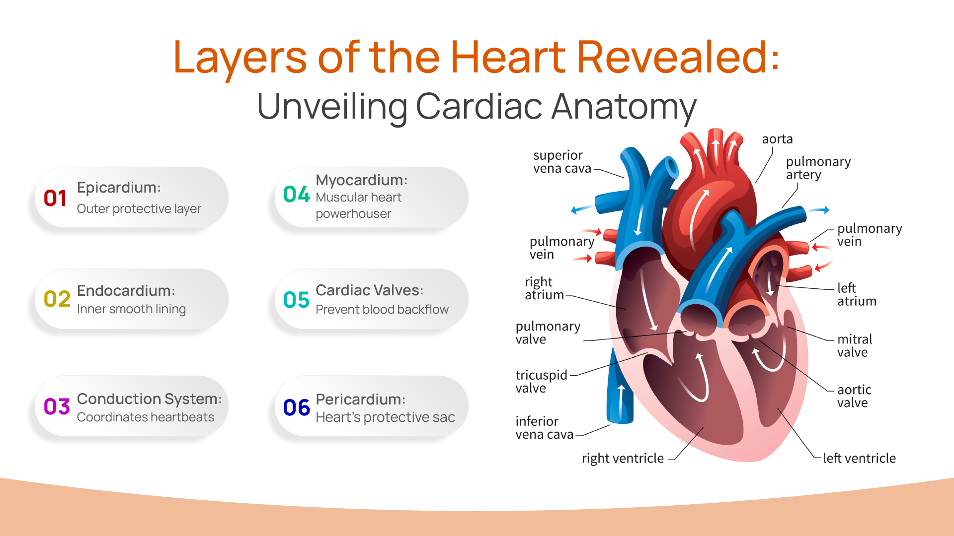
The different layers of the heart can be described by their distinct qualities. The outer layer of the heart The Epicardium is the protective layer. After this, the myocardium is the muscular powerhouse of the heart. The endocardium is the inner lining of the heart. The cardiac valves are the guardians of the blood flow. Finally, the conduction system of the heart is its electrical system contributing to the circulation of blood to vital parts of the body.
The Epicardium
The epicardium is the outermost layer of the heart. It is a thin layer of connective tissue that also contains fat, blood vessels, and lymphatics. The primary function of the epicardium is to provide a smooth, lubricated surface that allows the heart to move easily within the pericardial sac as it beats.
The epicardium is part of the serous pericardium, which is divided into two layers: the parietal layer lining the fibrous pericardium and the visceral layer, or epicardium, covering the heart. This double-layered structure creates a pericardial cavity filled with pericardial fluid, reducing friction during heart contractions.
The Myocardium
Beneath the epicardium lies the myocardium, the thick, muscular middle layer of the heart. This is the most substantial layer and is responsible for the contractile function of the heart. The myocardium is composed of cardiac muscle cells known as myocytes, which are unique in their ability to contract rhythmically and continuously without fatigue.
The myocytes are organized into a complex network of fibers that enable the heart to contract in a coordinated manner. These fibers are arranged in a spiral pattern, allowing for efficient pumping action. When the myocardium contracts, it generates the force needed to propel blood out of the heart and into the circulation. This muscular layer is particularly thick in the ventricles, especially the left ventricle, as it needs to generate high pressure to pump blood throughout the body.
The Endocardium
The endocardium is the innermost layer of the heart, lining the heart chambers and covering the heart valves. It is a thin, smooth layer composed of endothelial cells that provides a slick surface to facilitate smooth blood flow within the heart. This layer is crucial in maintaining a non-thrombogenic (non-clotting) surface, which is vital for preventing blood clots within the heart chambers.
The endocardium also plays a role in regulating the function of the myocardium by releasing certain signaling molecules. It is continuous with the endothelium of the blood vessels, creating a seamless transition between the heart and the vascular system. This continuity is critical for maintaining the integrity of the cardiovascular system.
The Cardiac Valves
Although not a distinct layer like the epicardium, myocardium, or endocardium, the cardiac valves are crucial components of the heart’s anatomy. The main function of these valves is to prevent the backflow of blood to the heart. These valves, composed of specialized tissue, ensure unidirectional blood flow through the chambers of the heart. There are four main valves in the heart:
- Tricuspid Valve: Located between the right atrium and right ventricle, this valve prevents the backflow of blood into the atrium during ventricular contraction.
- Pulmonary Valve: Situated between the right ventricle and the pulmonary artery, it opens to allow blood to flow into the lungs and closes to prevent backflow into the ventricle.
- Mitral Valve: Found between the left atrium and left ventricle, it ensures that blood flows from the atrium to the ventricle without regurgitating back.
- Aortic Valve: Located between the left ventricle and the aorta, it opens to permit blood to enter the systemic circulation and closes to prevent backflow into the ventricle.
Each valve’s precise function is vital for maintaining the efficiency of the heart’s pumping action and overall cardiovascular health.
The Cardiac Conduction System
Embedded within the layers of the heart, particularly the myocardium, is the conduction system of the heart. This specialized network of cells initiates and propagates electrical impulses that coordinate heartbeats. Key components of this system include the sinoatrial (SA) node, atrioventricular (AV) node, bundle of His, and Purkinje fibers.
- SA Node: Known as the natural pacemaker of the heart, it generates electrical impulses that set the heart rate.
- AV Node: Acts as a gateway that slows the electrical signal before it enters the ventricles, ensuring the atria have time to contract fully before the ventricles do.
- Bundle of His and Purkinje Fibers: These pathways distribute the electrical impulse throughout the ventricles, ensuring synchronized contraction of the ventricular muscles.
The Pericardium
Surrounding the heart is the pericardium, a double-walled sac that offers additional protection and support. The pericardium has two main layers:
- Fibrous Pericardium: The tough, outer layer that anchors the heart within the chest cavity and prevents overexpansion.
- Serous Pericardium: The inner layer, which is further divided into the parietal layer lining the fibrous pericardium and the visceral layer (epicardium) covering the heart.
The pericardial cavity, filled with pericardial fluid, lies between these layers, providing a lubricated environment that minimizes friction during heartbeats.
In conclusion, all the layers of the heart serve different purposes in the functioning of the heart. Without these layers, the heart would easily collapse and would not be able to perform its functions, which would cause chaos in the entire body. That is why it is important to take care of the layers of the heart and stay vigilant.