


Author:- Mr. Ritesh Sharma
A STEMI (ST-Elevation Myocardial Infarction) ECG is a critical diagnostic tool for detecting one of the most severe forms of heart attacks. Recognizing and interpreting a STEMI ECG promptly can be the difference between life and death. This blog will delve into the specifics of STEMI ECG, how it works, why it’s vital, and what healthcare professionals look for when diagnosing a STEMI.
What is a STEMI?
A STEMI is a type of heart attack characterized by a long interruption to the blood supply, caused by a complete blockage of a coronary artery. This blockage leads to significant damage to the heart muscle, as it deprives it of oxygen and nutrients. The term “STEMI” stands for ST-Elevation Myocardial Infarction, which indicates that the ST segment of the ECG is elevated. This elevation is a crucial indicator that a full-thickness heart muscle injury is occurring.
The Importance of Early Detection
Early detection of a STEMI is vital for several reasons. Firstly, the sooner a patient is diagnosed, the quicker they can receive treatment, which may include medications to dissolve the blockage, procedures to open the artery, or even surgery. Secondly, early intervention can significantly reduce the extent of heart muscle damage, improving the patient’s prognosis and reducing the risk of complications such as heart failure or cardiac arrhythmias.
Understanding the ECG
An Electrocardiogram (ECG or EKG) is a test that measures the electrical activity of the heart. During a heart attack, the heart’s electrical patterns change in ways that can be detected by an ECG. In a STEMI, the most telling sign is the elevation of the ST segment, which appears as a distinct pattern on the ECG reading.
Key Features of a STEMI ECG
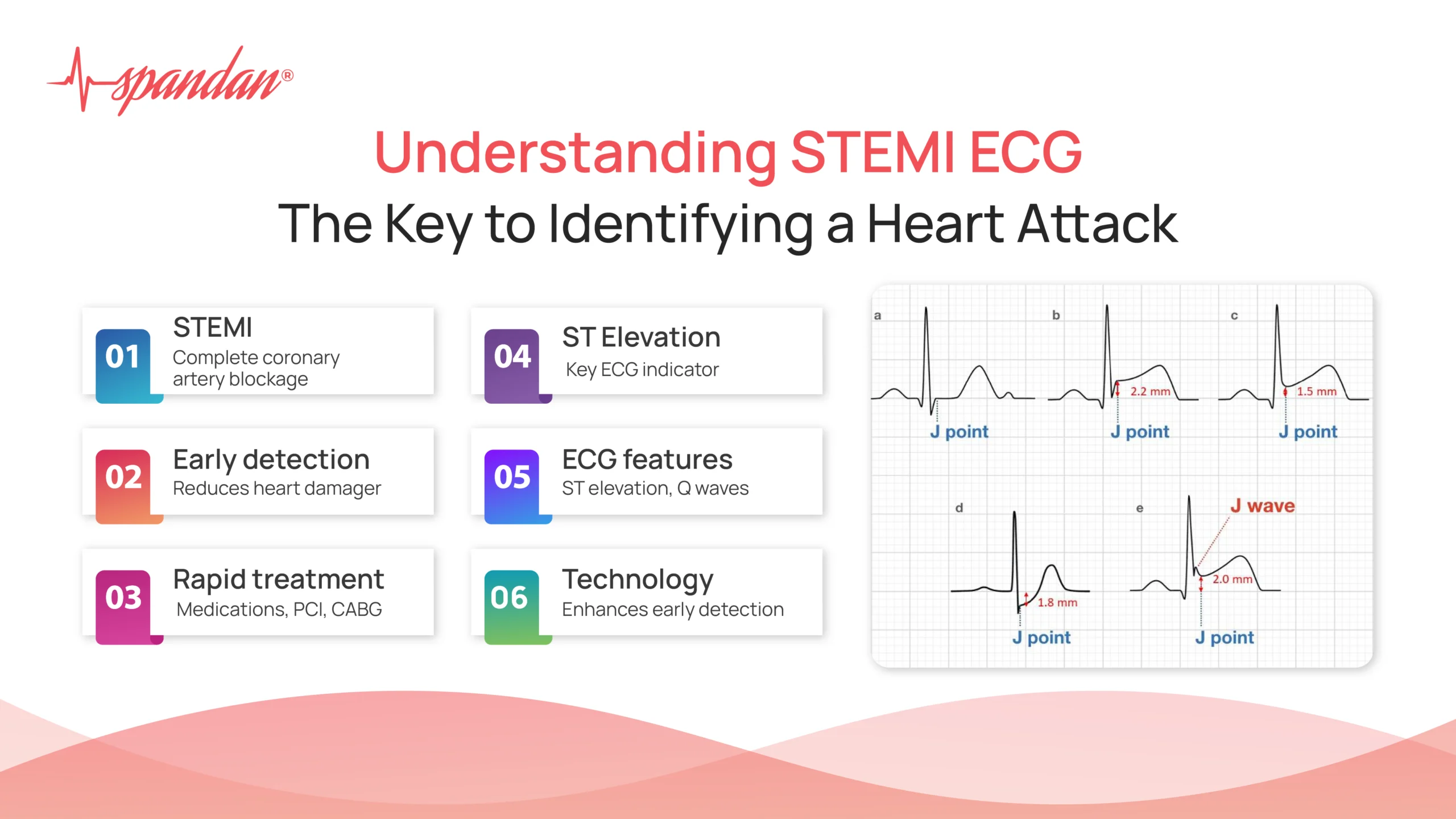
- ST Segment Elevation: The hallmark of a STEMI ECG is the elevation of the ST segment. This elevation must be at least 1 mm (0.1 mV) in two or more contiguous leads. In some cases, the elevation may be more pronounced.
- Reciprocal Changes: In addition to ST segment elevation, reciprocal changes may be observed in the leads opposite the site of infarction. These changes often present as ST segment depression, also known as ST depression.
- Q Waves: Pathological Q waves may develop hours to days after the infarction, indicating that a full-thickness myocardial infarction has occurred.
- T Wave Inversions: T wave inversions may also appear showcasing T-wave abnormalities, signifying ongoing ischemia or injury to the heart muscle.
Reading a STEMI ECG
Interpreting a STEMI ECG requires training and experience. Healthcare professionals look for several key indicators:
- Location of Elevation: The specific leads where ST elevation is observed can help determine the location of the infarction. For example, elevation in leads II, III, and aVF suggests an inferior wall infarction, while elevation in leads V1 to V4 indicates an anterior wall infarction.
- Magnitude of Elevation: The extent of ST elevation can provide clues about the severity of the infarction.
- Associated Symptoms: Clinical correlation is essential. Symptoms like chest pain, shortness of breath, sweating, and nausea, when combined with a STEMI ECG, strongly suggest a heart attack.
Treatment Protocols
Once a STEMI is identified via an ECG, immediate treatment is crucial. Treatment options include:
- Medications: Thrombolytic agents can dissolve the clot causing the blockage. Antiplatelet drugs and anticoagulants prevent further clot formation.
- Percutaneous Coronary Intervention (PCI): This is a non-surgical procedure that uses a catheter to place a stent and open up the blocked artery.
- Coronary Artery Bypass Grafting (CABG): In severe cases, surgery may be needed to bypass the blocked artery.
Time is a critical factor; the mantra “time is muscle” underscores the urgency of treating a STEMI to save heart muscle and improve outcomes.
Real-life Application of STEMI ECG
Consider a scenario where a 58-year-old male presents to the emergency department with severe chest pain, radiating to his left arm, and accompanied by sweating and shortness of breath. An ECG is promptly performed, revealing ST segment elevations in leads V2 to V5, indicating an anterior wall STEMI.
In this case, the immediate interpretation of the STEMI ECG allows for rapid decision-making. The patient is quickly moved to the cardiac catheterization lab for PCI, where the blockage is identified and a stent is placed, restoring blood flow to the heart. Post-procedure, the patient’s symptoms improve, and follow-up ECG shows resolution of the ST elevation, i.e. the abnormal ECG turns into a normal ECG.
This case illustrates the lifesaving potential of a timely STEMI ECG. The quick identification and treatment significantly reduce the extent of heart muscle damage and improve the patient’s prognosis.
The Role of Technology in STEMI ECG
Advancements in technology have further enhanced the efficacy of STEMI ECGs. Portable ECG devices and telemedicine allow for earlier detection, even before the patient reaches the hospital. For instance, paramedics can perform an ECG in the ambulance and transmit the results to the hospital, enabling the medical team to prepare for immediate intervention upon arrival.
The STEMI ECG is a vital tool in the early diagnosis and treatment of one of the most severe forms of heart attacks. By understanding the key features and interpretation of a STEMI ECG, healthcare professionals can make rapid and accurate decisions that significantly impact patient outcomes. As technology continues to advance, the potential for even earlier detection and treatment of STEMI will continue to improve, ultimately saving more lives.
In summary, the STEMI ECG is not just a diagnostic test; it is a lifesaving measure that underscores the importance of swift medical response in the face of a heart attack. Recognizing the signs, interpreting the results accurately, and acting promptly are crucial steps in the battle against heart disease.